بحث من تقديم الدكتور يوسف الشرف.. رئيس مركز الشرف التخصصي للعظام والعمود الفقري.. مملكة البحرين:
مرض القرص التنكسي هو حالة تسبب التدهور أو الكسر للأقراص الفقرية في العمود الفقري. الأقراص الفقرية هي منصات امتصاص الصدمات التي تقع بين الفقرات الخاصة بك. الفقرات هي سلسلة من العظام التي تشكل العمود الفقري الخاص بك. أمراض القرص التنكسية يمكن أن تحدث في أي جزء من العمود الفقري. وهي تتطور أكثر في كثير من الأحيان في أسفل الظهر. التقدم في العمر يمكن أن يجعل الأقراص تفقد السوائل، تنهار، وأحيانا تتمزق. وهذا يقلل من مساحة بين الفقرات، مما يجعل بعض الناس أقصر مع تقدمهم في السن. تدهور القرص يؤثر على هيكل الفقرات. هذه التغيرات يمكن أن تؤدي الى الضغط على الحبل الشوكي والأعصاب. معظم أعراض الألم يمكن علاجها بشكل غير جراحي. ومع ذلك، ينصح بإجراء الجراحة إذا كان العمود الفقري غير مستقر أو اذا كان الألم لا يمكن أن يزول من خلال وسائل أخرى.
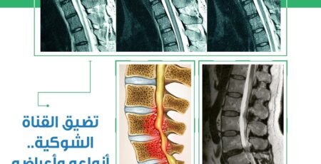
*تشريح: يتكون العمود الفقري من سلسلة من العظام تسمى الفقرات. هناك مناطق مختلفة في العمود الفقري، التي تعرف من انحنائها ووظيفتها. تحتوي رقبتك على العمود الفقري العنقي. و يتألف من سبع فقرات صغيرة. تحتوي منطقة الصدر والعمود الفقري الصدري، على 12 فقرة. يقع العمود الفقري القطني تحت الخصر. و يحتوي على خمسة فقرات كبيرة. وتندمج باقي فقرات العمود الفقري في تشكيل الورك والحوض . الجزء الخلفي من كل فقرة ينحني لتشكيل الصفيحة. الصفيحة تكون غطاء مثل السقف تغطي الفراغ في كل فقرة. الفراغ في وسط كل فقرة يشكل القناة الشوكية. الحبل الشوكي والأعصاب، والشرايين تعبر خلال قناة النخاع الشوكي. الحبل الشوكي والأعصاب يقومون بإرسال الرسائل بين الجسم والدماغ. تقع الأقراص الفقرية ما بين العنق والصدر، والفقرات القطنية. تتكون الأقراص من نسيج ضام قوي. وتسمى الطبقة الخارجية الخشنة بالحلقة الليفية. ويطلق على مركز المادة الهلامية النواة اللبية. يحتوي القرص الصحي على حوالي 80٪ من الماء. الأقراص واثنين من صفائح العمود الفقري يربطون الفقرات ببعضها البعض. الأقراص والمفاصل يسمحون بالحركة وتوفير الاستقرار. تعمل الأقراص أيضا بمثابة وسادة لامتصاص الصدمات لحماية الفقرات.
*الأسباب: مع تقدمنا في العمر، تفقد الأقراص محتوى الماء فتصبح أقصر وأقل مرونة. وبمجرد إصابة الأقراص، يقل وصول الدم إليهم فتتدهور. بدون القرص الواقي، يصبح العمود الفقري غير مستقر من الناحية الهيكلية وغير قادر على تحمل الإجهاد، وهذا قد يؤدي إلى مشاكل أخرى. الأقراص الفقرية بمثابة وسادة بين الفقرات فعندما ينحل القرص، يمكن أن تحتك العظام ببعضها. قد يحدث نمو غير طبيعي للعظام يسمى بالزائدة العظمية، يمكن أن تنمو في المفصل وتدخل القناة الشوكية. فتسبب الألم والتورم وتعطل الحركة. قد تحدث تغييرات في بنية العمود الفقري يمكن أن تسبب إنزلاق فقرة واحدة إلى الأمام والخروج من مكانها، وهي حالة تسمى الانزلاق الفقاري. بدون القرص ، الأربطة والمفاصل في الفقرات قد تكبر للمساعدة في تعويض الضغط على العمود الفقري و قد تمتد إلى القناة الشوكية فتسبب تضييق. عندما تضيق القناة الشوكية يمكن أن تضغط على الحبل الشوكي والأعصاب، مما يؤدي إلى ألم وفقدان في الوظيفة، وهي حالة تسمى تضيق العمود الفقري.
مرض القرص التنكسي يمكن أن يؤدي أيضا إلى انزلاق غضروفي. طبقة القرص الخارجي، والحلقة، يمكن تنشق أو تتمزق تحت الضغط. يحدث الانزلاق الغضروفي عندما ينشق الطوق ويخرج المحتوى الداخلي و النواة اللبية من القرص. عندما تخرج المحتويات الداخلية تتصل مع الأعصاب في العمود الفقري فتؤدي الى تهيجها وانتفاخها ، مما يؤدي إلى الألم.
يمكن أن تحدث الأمراض التنكسية للقرص في أي منطقة من العمود الفقري. ويحدث ذلك غالبا في منطقة أسفل الظهر. الأطباء ليسوا متأكدين من السبب الدقيق لهذا المرض. ويبدو أن الشيخوخة، والصدمات النفسية، والتهاب المفاصل تسهم في هذه الحالة. ويعتقد الأطباء أن العوامل الوراثية والبيئية، والمناعة الذاتية تلعب دورا في ذلك. بالإضافة إلى ذلك، عوامل نمط الحياة، بما في ذلك التدخين أو الأنشطة المتكررة مضنية، مثل الجمباز ورفع الأثقال يمكن أن تسهم في تنكس القرص كذلك. الأمراض التنكسية للقرص تحدث معظم الأحيان لمن هم في اواسط العمرأوالبالغين.
*الأعراض: الأمراض التنكسية للقرص قد أو قد لا تسبب أعراضا. إذا كان لديك أعراض، قد تشعر بأنواع مختلفة من الألم في الظهر أو الرقبة. قد تشعر بألم مفاجئ بعد إصابة أو قد يبدأ الألم تدريجيا ويزيد مع مرور الوقت. قد يكون الألم قويا لدرجة أنه يعيق نشاطاتك اليومية. قد تشعر بألم حارق، وضغط، وخدر، أو وخز. الجلوس قد جعل الأعراض تزيد ، في حين الاستلقاء قد يساعد على تخفيف الألم. تبعا للمكان الذي يقع فيه القرص المتنكس في العمود الفقري ، قد تتأثر ذراعيك أو الساقين أيضا. في حالات نادرة، فقدان السيطرة على المثانة والأمعاء يرافقه ضعف كبير في الذراع والساق يشير إلى وجود مشكلة خطيرة محتملة. في هذه الحالة النادرة، يجب عليك الحصول على عناية طبية فورية.
*التشخيص: طبيبك يمكن تشخيص القرص التنكسي عن طريق إجراء الفحص البدني و الصور التشخيصية. سوف يسألك طبيبك عن الأعراض والتاريخ الطبي. سوف يطلب منك القيام بحركات بسيطة للمساعدة على تقييم قوتك العضلية وحركة المفاصل، والاستقرار. بما إن الأعصاب ممتدة من العمود الفقري الى الجسم، سيقوم الطبيب بإجراء فحص للجهاز العصبي لذراعيك و الساقين ليتحقق من وظيفة الأعصاب.
طبيبك سوف يأمرك بعمل الأشعة السينية لمعرفة حالة الفقرات في العمود الفقري. أحيانا الأطباء يقومون بحقن صبغة في العمود الفقري لتعزيز صور الأشعة السينية في إجراء يسمى تصوير النخاع. تصوير النخاع يمكن أن يشير إلى ما إذا كان هناك ضغط على الحبل الشوكي أو الأعصاب من فتق الأقراص، الزوائد العظمية ، أو الأورام.
قد يأمر الطبيب أيضا بعمل التصوير المقطعي أو التصوير بالرنين المغناطيسي للحصول على صورة أفضل للعمود الفقري . تظهر الاشعة المقطعية شكل وحجم القناة الشوكية والهياكل حولها. والاشعة المقطعية مفيدة لتحديد أي قرص تالف. طبيبك قد يحقن صبغة في القرص في إجراء يسمى صورة القرص. يوفر صورة القرص إطلالة على الهيكل الداخلي للقرص ويمكن أن يساعد في تحديد ما اذا كان هو مصدر الألم. وعادة ما تليها مباشرة الاشعة المقطعية. التصوير بالرنين المغناطيسي حساس جدا. لأنه يوفر صورا أكثر تفصيلا عن الأقراص والأربطة والحبل الشوكي والجذور العصبية، أو الأورام. الأشعة السينية، الأشعة المقطعية، والرنين المغناطيسي هي إجراءات غيرمؤلمة.
*العلاج: معظم المصابين بالأمراض التنكسية للقرص يمكن علاجهم بالطرق غير الجراحية التي تهدف إلى تخفيف الآلام وتعديل النشاط. بعض الوصفات الدوائية يمكن ان تستخدم لتخفيف الألم. إذا كانت الأعراض لم تتحسن بشكل كبير مع هذه الأدوية، فإن طبيبك قد يحقن المفصل مع دواء كورتيكوستيرويد. دواء كورتيكوستيرويد هو مخلص للآلام وآمن نسبيا.
قد يوصي طبيبك بالراحة وارتداء حزام الظهر أو الرقبة. أخصائي العلاج الطبيعي أو المهني يمكن أن يوفر علاجات للحد من الألم، وتشنجات العضلات، والتورم. وسيقوم بتعليمك تمارين لتقوية عضلات ظهرك أو الرقبة.
*العملية الجراحية: العلاجات غير الجراحية للأمراض التنكسية للقرص يقومون بتخفيف الألم واستعادة الوظيفة، لكنهم لا يستطيعون تصحيح التشوهات الهيكلية، مثل ضيق القناة الشوكية. ينصح بالجراحة عندما تفشل العلاجات غير الجراحية من تحسين الأعراض. وينصح بالجراحة إذا كان القرص يضغط مباشرة على العصب أو الحبل الشوكي، مما يسبب إعاقة كبيرة في الوظائف. وكذلك إذا أصبح الضعف في الساق يسوء تدريجيا أو إذا كان هناك مشاكل مرتبطة بالمثانة والأمعاء.
نوع الجراحة تعتمد على موقع القرص التنكسي في العمود الفقري ومدى الحالة. دمج العمود الفقري هو نوع من الجراحة الأكثر استخداما للأمراض التنكسية للقرص. تتضمن إزالة القرص التنكسي ودمج اثنين أو أكثر من الفقرات معا لوقف الحركة وتخفيف الألم الناجم عن الحركة. هناك تقنيات مختلفة لعملية الدمج في العمود الفقري تعتمد على المنطقة المصابة.
سوف يتم تخديرك للعملية الجراحية. سيقوم الطبيب بإجراء الجراحة من الأمام أو الخلف من العمود الفقري ، اعتمادا على المنطقة المتأثرة من العمود الفقري. للجراحة الخلفية المستخدمة في منطقة أسفل الظهر، سوف يقوم الجراح بإجراء شق في منتصف العمود الفقري. للجراحة الأمامية المستخدمة في منطقة العنق ، سوف يقوم الجراح بإجراء شق في منتصف العنق. سيتم إبعاد العضلات وغيرها من الهياكل جانبا بعناية للسماح بالوصول إلى الفقرات. سوف يقوم الجراح بإزالة كل أو جزء من القرص التنكسي.
بعد ذلك، الجراح يضع قطعة من العظم في مساحة القرص الفارغ. ويتكون من شرائح صغيرة من العظم مأخوذة من الورك أثناء الجراحة. العظم المزروع أحيانا يستخدم من متبرع. يتم وضع العظم بين الفقرات. يتم تثبيت العظمة بالمسامير والصفائح في الرقبة، وبالمسامير والقضبان في العمود الفقري القطني ، لدمج الفقرات معا. عند الانتهاء من الدمج في العمود الفقري ، سوف يقوم الجراح بإغلاق الشق مع الغرز. سوف تتلقى الادوية المسكنة للألم مباشرة بعد الجراحة. سوف تقوم بارتداء حزام الظهر أو الرقبة ، اعتمادا على موقع الجراحة، الى حين التئآم الدمج.
*الشفاء: عملية الشفاء تعتمد على نوع الجراحة ومنطقة العمود الفقري المتضررة ، ومدى حالتك. عموما، وقت الشفاء للدمج في منطقة العنق من أربعة إلى ستة أسابيع. الدمج في العمود الفقري في المنطقة القطنية يستغرق وقت اطول في الشفاء.
يجب أن تتوقع أن تبقى بين عشية وضحاها أو بضعة أيام في المستشفى، وهذا يتوقف على نوع وموقع الجراحة. قد تحتاج إلى القليل من المساعدة من شخص آخر خلال الأيام القليلة الأولى أو أسابيع في المنزل. إذا لم يكن لديك أفراد من العائلة أو صديق قريب، تحدث مع طبيبك حول الترتيبات البديلة الممكنة.
طبيبك سوف يقيد في البداية مستوى النشاط الخاص بك وتحديد وضعيات للجسم. يجب تجنب رفع الاثقال ،الأعمال المنزلية، و العمل إلى ان يسمح لك الطبيب. قم بارتداء حزام الظهرأو الرقبة للحصول على الدعم. يمكنك ان تزيد تدريجيا مستوى نشاطك. عندما يلتئم الاندماج ، أخصائي العلاج الطبيعي سيقوم بتعليمك تمارين التقوية و ميكانيكا الجسم، الوضعيات المناسبة لعمودك الفقري خلال الوقوف، الجلوس، والنوم، ورفع الأشياء.
*الوقاية: من المهم أن تلتزم القيود وممارسة التمارين عند العودة إلى البيت. يجب عليك استخدام ميكانيكا الجسم السليم خلال جميع الأنشطة. لا تدخن. التدخين يزيد من خطر المضاعفات الجراحية ويعيق اندماج العظام.
Degenerative Disc Disease – Spine Degeneration
by dr. yousif sharaf
Degenerative Disc Disease is a condition that causes the intervertebral discs in the spine to deteriorate or break down. Intervertebral discs are the shock-absorbing pads located between your vertebrae. The vertebrae are the series of bones that make up your spine. Degenerative Disc Disease can occur in any part of the spine. It develops more frequently in the lower back. Aging can cause the discs to lose fluid, collapse and sometimes rupture. This decreases the space between the vertebrae, which is why some people become shorter as they age. As the disc deteriorates, it affects the structure of the vertebrae. These changes can lead to conditions that put pressure on the spinal cord and nerves. Most symptoms of pain can be treated non-surgically. However, surgery is recommended if the spine is unstable or when pain cannot be relieved by other means. Play Video
Anatomy The spine is made up of a series of bones called vertebrae. There are different areas of the spine, defined by their curvature and function. Your neck contains the cervical spine. It is composed of seven small vertebrae. Your chest area contains the thoracic spine, with 12 vertebrae. The lumbar spine is located at and below your waist. It contains five large vertebrae. The remainder of the lower vertebrae in the spine are fused or shaped differently in formation with your hip and pelvis bones.
The back part of each vertebra arches to form the lamina. The lamina creates a roof-like cover over the back opening in each vertebra. The opening in the center of each vertebra forms the spinal canal. Your spinal cord, nerves, and arteries travel through the protective spinal canal. The spinal cord and nerves send messages between your body and brain.
Intervertebral discs are located in between the cervical, thoracic, and lumbar vertebrae. The discs are made up of strong connective tissue. Their tough outer layer is called the annulus fibrosus. Their gel-like center is called the nucleus pulposus. A healthy disc contains about 80% water.
The discs and two small spinal facet joints connect one vertebra to the next. The discs and joints allow movement and provide stability. The discs also act as a shock-absorbing cushion to protect the vertebrae.
Causes As we age, our discs lose water content. Our discs become shorter and less flexible. Once the discs are injured, they do not have the blood supply to repair themselves and they deteriorate. Without the protective disc, the spine can become structurally unstable and unable to tolerate stress, which may lead to other conditions.
Normally, the intervertebral discs act as a cushion between the vertebrae. When a disc degenerates, painful bone on bone rubbing can occur. Abnormal bone growths, called spurs or osteophytes, can grow in the joint and enter the spinal canal. The bone spurs add to pain and swelling, while disrupting movement. The changes in spine structure can cause one vertebra to shift forward and out of place, a condition called Spondylolisthesis.
Without the disc to act as a cushion, the ligaments and facet joints on the vertebrae may enlarge to help compensate for the stress on the spine. The overgrowth can extend into the spinal canal causing it to narrow. The narrowed spinal canal can compress the spinal cord and nerves, resulting in pain and loss of function, a condition called Spinal Stenosis.
Degenerative Disc disease can also lead to a herniated disc. The outer disc layer, the annulus, can tear or rupture under stress. A herniated disc occurs when the annulus ruptures and the inner contents, the nucleus pulposus, comes out of the disc. When the inner contents come in contact with the spinal nerves they become irritated and swell, resulting in pain.
Degenerative Disc Disease can occur in any region of the spine. It most commonly occurs in the lumbar area. Doctors are not sure of the exact cause of Degenerative Disc Disease. It appears that the aging process, trauma, and arthritis contribute to the condition. Doctors suspect that genetic, environmental, and autoimmune factors play a role. Additionally, lifestyle factors, including smoking or strenuous repetitive activities, such as gymnastics or lifting, may contribute to disc degeneration as well. Degenerative Disc Disease develops most frequently in middle aged or young adults with active lifestyles.
SymptomsDegenerative Disc Disease may or may not cause symptoms. If you have symptoms, you may feel various types of pain in your back or neck. You may experience sudden pain after an injury or your pain may start gradually and increase over time. Your pain may be so intense that it interferes with your daily activities. You may feel burning pain, pressure, numbness, or tingling. Sitting may make your symptoms increase, whereas lying down may help to relieve pain. Depending on where your degenerative disc is located in your spine, your arms or legs may be affected as well. In rare cases, the loss of bowel and bladder control accompanied by significant arm and leg weakness indicates a possible serious problem. In this rare case, you should seek immediate medical attention.
Diagnosis Your doctor can diagnose a degenerative disc by performing a physical examination and viewing medical images. Your doctor will ask you about your symptoms and medical history. You will be asked to perform simple movements to help your doctor assess your muscle strength, joint motion, and stability. Since the nerves from the spine travel to the body, your doctor will perform a neurological examination of your arms and or legs to see how the nerves are functioning.
Your doctor will order X-rays to see the condition of the vertebrae in your spine. Sometimes doctors inject dye into the spinal column to enhance the X-ray images in a procedure called a myelogram. A myelogram can indicate if there is pressure on your spinal cord or nerves from herniated discs, bone spurs, or tumors.
Your doctor may also order Computed Tomography (CT) scans or Magnetic Resonance Imaging (MRI) scans to get a better view of your spinal structures. CT scans provide a view in layers, like the slices that make up a loaf of bread. The CT scan shows the shape and size of your spinal canal and the structures in and around it. A CT scan is useful for determining which disc is damaged. Your doctor may inject dye into the disc in a procedure called a discogram. A discogram provides a view of the internal structure of a disc and can help to identify if it is a source of pain. It is usually immediately followed by a CT scan. The MRI scan is very sensitive. It provides the most detailed images of the discs, ligaments, spinal cord, nerve roots, or tumors. X-rays, myelograms, CT scans, and MRI scans are painless procedures.
Treatment Most people with Degenerative Disc Disease can be treated with non-surgical methods aimed at pain relief and activity modification. Over-the-counter medication or prescription medication may be used to ease your pain. If your symptoms do not improve significantly with these medications, your doctor may inject your joint with corticosteroid medication. Corticosteroid medication is a relatively safe pain reliever.
Your doctor may recommend that you rest and wear a back or neck brace. Occupational or physical therapists can provide treatments to reduce your pain, muscle spasms, and swelling. The therapists will also show you exercises to strengthen your back or neck muscles.
Surgery Non-surgical treatments for Degenerative Disc Disease are designed to relieve pain and restore function, but they can not correct structural deformities, such as narrowing of the spinal canal. Surgery is recommended when non-surgical treatments have provided minimal or no improvement of your symptoms. Surgery is advised if the disc is pressing directly on a nerve or the spinal cord, causing considerable loss of function. Surgery is also advised if your leg weakness becomes progressively worse or if you experience associated bladder and bowel problems.
The type of surgery that you have will depend on the location of the degenerative disc in your spine and the extent of your condition. Spinal Fusion is the type of surgery most frequently used for degenerative disc disease. Spinal Fusion involves removing the degenerative disc and fusing or securing two or more vertebrae together to stop movement and relieve pain caused by movement. There are various techniques for spinal fusion surgery and the area of the spine that is involved determines the approach. Your doctor will let you know what to expect.
You will be sedated for the surgery. Your doctor will perform the surgery from the front or back of your spine, depending on the region of the spine that is affected. For a posterior surgery used for the lumbar region, your surgeon will make an incision on the middle of your spine. For an anterior surgery used for the cervical area, your surgeon will make an incision on the middle of your neck. Your muscles and other structures will be moved aside with care to allow access to the vertebrae. Your surgeon will remove all or part of the degenerative or herniated disc. Next, the surgeon places a bone graft or interbody fusion cage in the empty disc space. A bone graft consists of small strips of bone taken from your hip during surgery. Sometimes allograft bone from a donor is used. The bone grafts are placed between the vertebrae. An interbody fusion cage may be used. It is a small container filled with bone shavings and placed between the vertebrae. The bone grafts or interbody fusion cage are surgically secured to the spinal column with surgical hardware, such as screws and plates in the neck, and screws and rods in the lumbar spine. The surgical hardware secures the vertebrae together and allows the bone grafts to heal, fusing together the vertebrae.
At the completion of your spinal fusion, your surgeon will close your incision with stitches. You will receive pain medication immediately following your surgery. You will wear a back or neck brace, depending on the location of your surgery, while your fusion heals.
Recovery The recovery process is different for everyone. It depends on the type of surgery that you had, the area of your spine affected, and the extent of your condition. Generally, the recovery time for a fusion in the cervical area is four to six weeks. Spinal fusion in the lumbar area has a longer recovery time. Your surgeon will let you know what to expect.
You should expect to stay overnight or a few days in the hospital, depending on the type and location of your surgery. You may need a little help from another person during the first few days or weeks at home. If you do not have family members or a friend nearby, talk to your doctor about possible alternative arrangements.
Your doctor will initially restrict your activity level and body positioning. You should avoid lifting, housework, and yard-work until your doctor gives you the okay to do so. You will wear a neck or back brace for support. You will gradually increase your activity level. Once your fusion has healed, physical therapists will teach you strengthening exercises. You will also learn body mechanics, proper postures for your spine, to use when you stand, sit, sleep, and lift objects.
Prevention It is important that you adhere to your restrictions and exercise program when you return home. You should use proper body mechanics during all activities. Do not smoke. Smoking increases the risk of surgical complications and hinders bone fusing.
X-ray Lumbar spine AP,Lateral : A human and postoperative treatment for degenerative lumbar disc disease by decompression and fix by iron rod and screws.Blue tone.


;)
اترك تعليقاً